Dr. Panagiotis A. Paraschakis
Interventional Neuroradiologist
Interventional neuroradiology (INR) or endovascular neurosurgery is the subspecialty that deals with the treatment of vascular diseases of the brain and spinal cord. Using intravascular catheters and image guidance, we perform a variety of diagnostic and therapeutic procedures
Imaging requires high-tech equipment to guide the instruments used during the procedure. Thin tubes (catheters) and wires are inserted into the blood vessels and guided to the affected area. Treatments, in the case of aneurysms or blood clot removal, are provided through these catheters. Diagnostic angiography is the foundation of the field, on which invasive procedures are based and performed to treat a variety of diseases.
The benefits that patients derive from this procedure are very significant. The main advantage is the reduction of pain but also the limited duration of hospitalization and recovery period, due to the almost bloodless procedure. Also, the use of advanced technology equipment ensures the precise localization of the affected area and therefore, the best treatment of the problem.
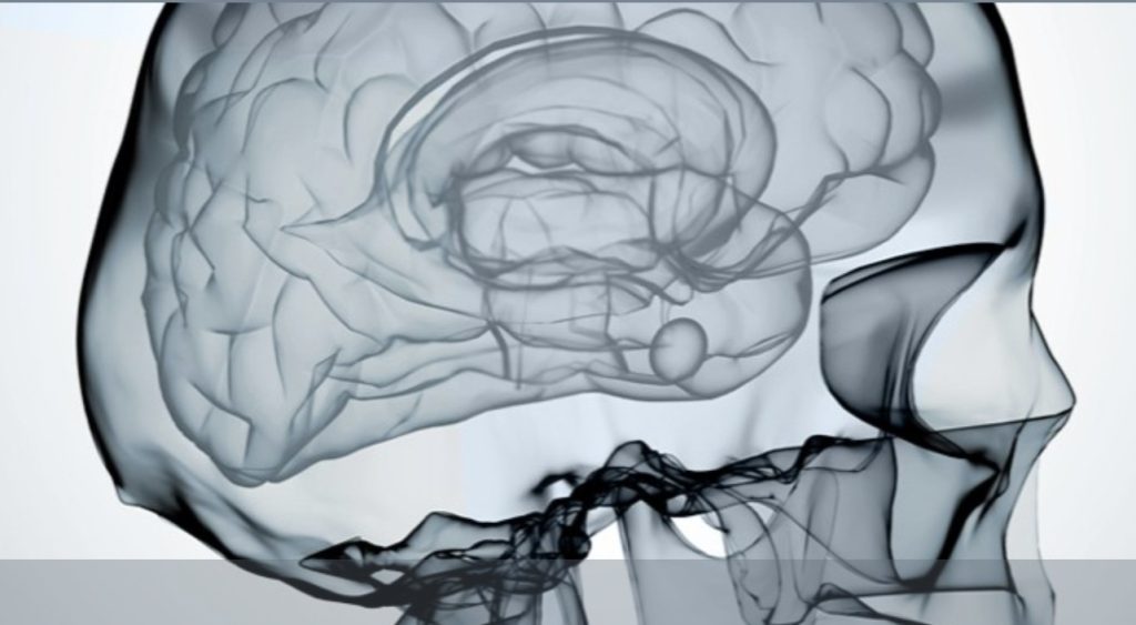
What vascular pathologies do we treat?
We deal with the treatment of brain aneurysms, ischemic strokes, arteriovenous malformations or communications of the brain and spinal cord, stenosis of the arteries of the brain and neck, chronic subdural hematomas, stenosis of the venous sinuses of the brain.
With what technique can these specific diseases be treated?
With minimally invasive methods, without open surgery, starting from the femoral artery and navigating catheters (tubes) and microcatheters, with the help of an X-ray machine, we reach the level of the lesion, which is most often located within the brain. There, with a variety of materials at our disposal, we can isolate a pathological communication, occlude an aneurysm, open a blocked vessel. The operation is performed under general anesthesia and most often requires a short hospitalization. The recovery is short and the patient returns to his daily life without the risk of bleeding.
Digital Brain Angiography
1
2
3
Brain Aneurysm
What is a brain aneurysm?
An intracranial aneurysm or cerebral aneurysm is a pathological swelling of the vessel wall that creates a sac. Inside this sac, blood circulates under pressure, which increases its fragility. The risk is its rupture and the occurrence of bleeding, which most often concentrates in the coverings of the brain, the meninges.
Known risk factors for aneurysms include, in addition to heredity, smoking, hypertension, and excessive alcohol consumption.
How are aneurysms discovered?
Often aneurysms are found incidentally during a test (CT or MR angiography) performed for another reason. In other cases, they are discovered after they rupture, which causes a sudden and severe headache that may be accompanied by vomiting or even neck pain.
How are aneurysms treated?
There are two types of therapeutic approach to aneurysms.
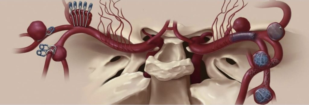
The neurosurgical approach, where the aneurysm is approached with an open operation and isolated from the circulation with one or more clips.
And the endovascular approach. In this approach, we navigate the materials through the arteries and place metal coils inside the aneurysm, alone or in combination with a stent, or we place a special type of flow-modifying stent in the artery carrying the aneurysm.
All aneurysms need therapy?
Aneurysms that have bled require emergency treatment. For aneurysms that are found incidentally, we assess their risk on a case-by-case basis, depending on their size, shape, location, and the presence of risk factors, and recommend treatment or monitoring.
Aneurysm | Clinical Cases
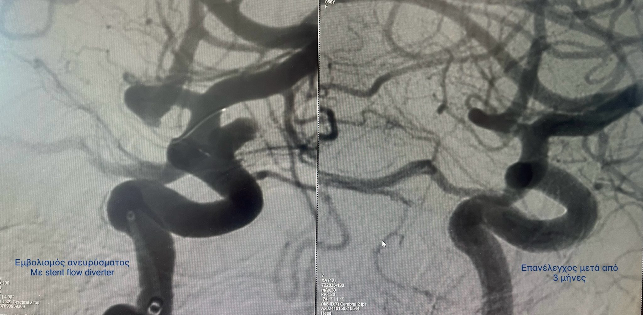
Embolization of an internal carotid aneurysm with the stent flow diverter technique
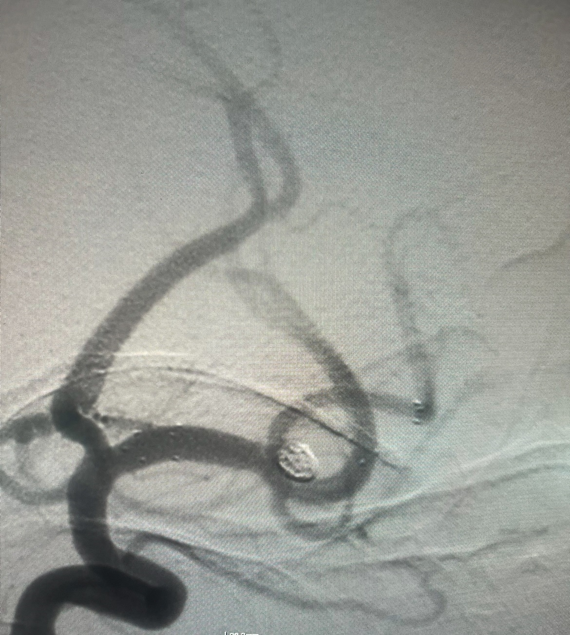
Embolization of an aneurysm of the left middle cerebral artery
Embolization of ruptured aneurysm with coils
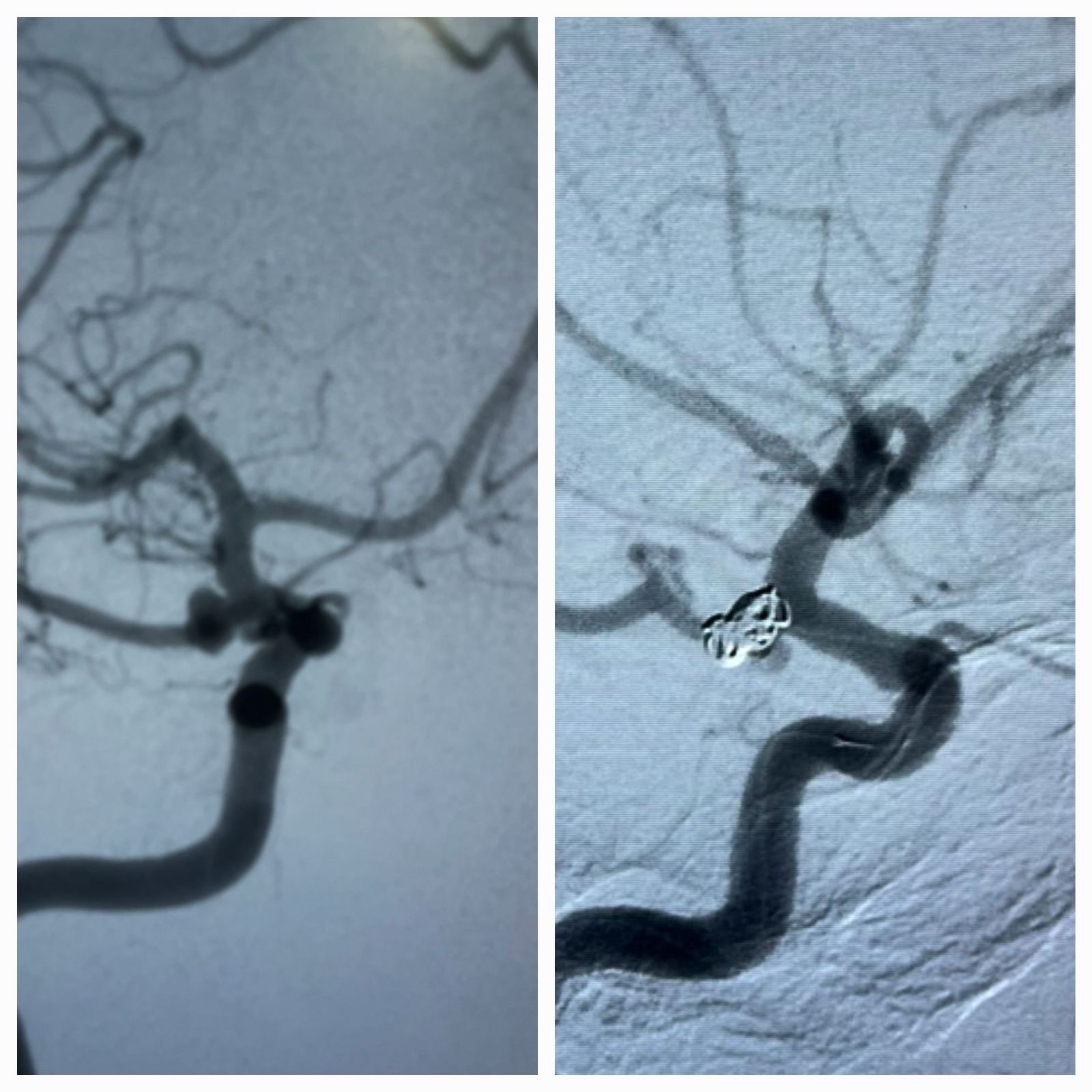
Anterior anastomotic artery aneurysm. Embolization with endovascular stent and coils
Ischemic Stroke (Stroke)
Ischemic stroke is the leading cause of disability in the Western world. It is caused by the blockage of an artery in the brain by a clot that most often originates in the heart or carotid arteries.
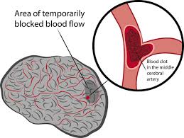
What are the symptoms?
Symptoms such as facial paralysis with drooping of the corner of the mouth, loss of strength or paralysis of the arm and leg on the same side, difficulty or weakness of speech or understanding, loss of vision, loss of balance, instability, numbness or loss of sense of touch, may be symptoms of a stroke.
What are the movements that should be made?
In the event that we witness such an episode in a relative, friend or random person, we must immediately call an ambulance so that the patient can be immediately transported to the hospital. There, he will be evaluated by specialists and referred for further tests, such as a brain scan (CT, CT angiography). In the event that a blockage of an important (large vessel) that supplies the brain is found, the patient needs to undergo a therapeutic operation.
What is the treatment for Ischemic Stroke?
There are two main therapeutic approaches that we follow in the case that the conditions are met. First, the intravenous administration of a drug that can dissolve the clot (intravenous thrombolysis). However, the invasive removal of the clot is more effective. The operation is called mechanical thrombectomy. The faster it takes place, the greater the chances that the patient will be autonomous.
Which risk factors can be modified?
After an ischemic stroke, whether it has significant consequences for the patient or not, we must perform a complete cardiac examination, to exclude or treat cardiac pathologies that can lead to a new episode. The vessels that supply the brain (the carotid and vertebral arteries) must also be checked, as well as the large intracranial vessels (internal carotid, middle cerebral arteries, vertebral, basilar artery).
Is arterial stenosis treated?
In the case where a stenosis in the arteries, before or within the brain, is symptomatic (causing ischemic stroke), it is highly likely that treatment is needed. This can be done endovascularly, where at the point of stenosis we open the artery with a balloon (angioplasty) and place a stent.
Arteriovenous Malformation (AVM)
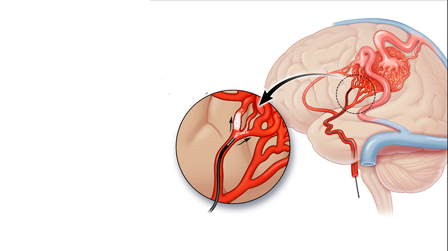
It is a pathological form of communication of arteries with veins, mediated by dysplastic vessels (nucleus/nidus). This pathology can be found incidentally (after MRI or CT scan), after an epileptic episode or after hemorrhage (cerebral parenchymal hematoma).
Frequently Asked Questions
On the Arteriovenous Malformation (AVM).
What is the strategy that we follow in arteriovenous communications?
When we find an arteriovenous malformation by chance, the next step is to perform a digital brain angiography (DSA brain) to identify its exact characteristics and risk. If we find the additional presence of aneurysms or arteriovenous malformations, treatment is probably needed. Otherwise, the usual tactic is to monitor the AVM.
If the AVM bleeds, treatment is needed within 4 to 6 weeks after the episode.
How is an arteriovenous malformation (AVM) treated today?
There are three different approaches to the AVM.
Intravascularly, with technique through the arteries or the veins with microtubes we get to the point of pathology and with a special liquid embolic material we occlude pathology.
With an open neurosurgical approach where externally approached the pathology and isolated with the use of clips or cauterization.
Third way is by the use of usually a single dose of radiation (gamma-knife/cyber knife). This method works progressively in 3 to 5 years.
You may need a combination of the methods of treatment depending on the case.
Arteriovenous Communication (Fistula)
This pathology is characterized by direct communication of an artery or arteries with veins. AVC or fistula can be found either incidentally or after a hemorrhagic episode. In the first case, we perform a digital cerebral angiography, where we classify the fistula and assess the bleeding risk. If we judge that it is high, we recommend surgery to the patient using the same techniques. In the case of hemorrhage, the surgery is performed in the acute phase.
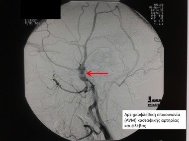
Where is arteriovenous malformation or arteriovenous communication located?
These diseases are located throughout the body. The object of study and treatment of neuroradiology are these vascular pathologies of the brain (intracranial) and the spinal cord. Their localization in the spinal cord is characterized by progressive paralysis of the lower extremities.
Arteriovenous Fistula | Clinical Cases
Arteriovenous communication embolism
Chronic Subdural Hematoma
This pathology mainly concerns patients over 70 years of age and concerns episodes of blood collection in the coverings of the brain, specifically under the dura mater. Usually, these are repeated episodes with a wide clinical presentation, from lack of symptoms to paralysis and cognitive disorders.
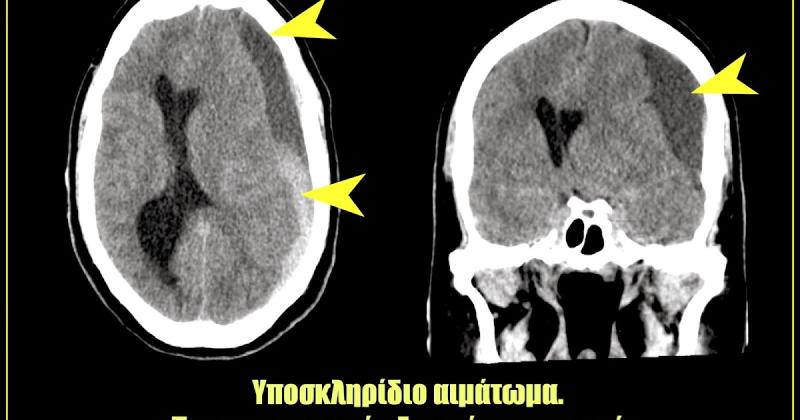
What is the treatment for chronic subdural hematoma?
Treatment can be observation or surgical drainage. Recently, a new technique has been introduced, the embolization of the middle meningeal artery, which has been shown to have very positive results in terms of accelerating the time needed for hematoma absorption, as well as reducing recurrent episodes. The result is that no additional surgical procedures are needed. Embolization can be done in combination with surgical removal of the hematoma, but also as the only treatment.
Collaborating Hospitals
Hospitals
Communication
Send us an email for more information or to evaluate your exams.

Dr. Panagiotis A. Paraschakis
Interventional Neuroradiologist
Dr Panagiotis A. Paraschakis is a graduate of the Department of Medicine of the Aristotle University of Thessaloniki, Greece. He obtained his specialization in Radiodiagnostics in 2016 in Athens.
He specialized in interventional neuroradiology at the Fondation Adolphe de Rothschild hospital in Paris, France. He worked as a specialist at the Hôpital Neurologique in Lyon and Hôpital Pasteur II in Nice, France.
Since May 2021, he has been working in Cyprus.
Since July 2025, he has been an instructor in interventional neuroradiology at the Alexandroupolis General University Hospital.